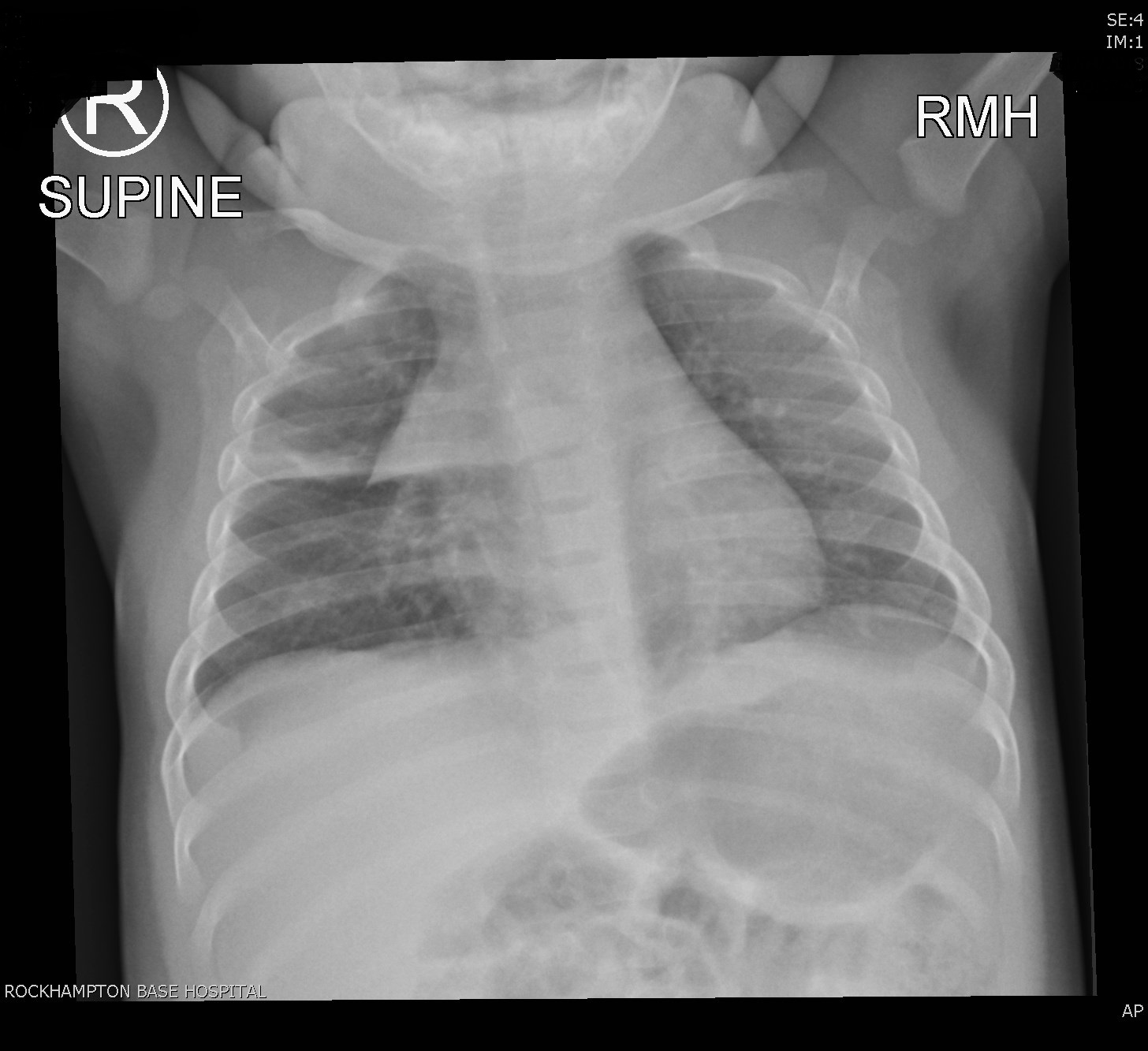
FINDINGS
Normal cardiomediastinal contour. Note the normal appearing thymic shadow.
Lateral to this in the right lung mid zone is patchy atelectasis and consolidation involving the right upper lobe. This is likely infectious in nature. Remaining lungs and pleura are clear.
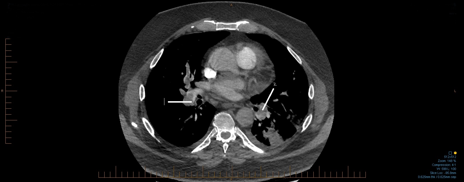
Suboptimal opacification of the pulmonary arteries.With this in mind, there are filling defects extending into the right
and left main pulmonary arteries in keeping with extensive central pulmonary emboli.

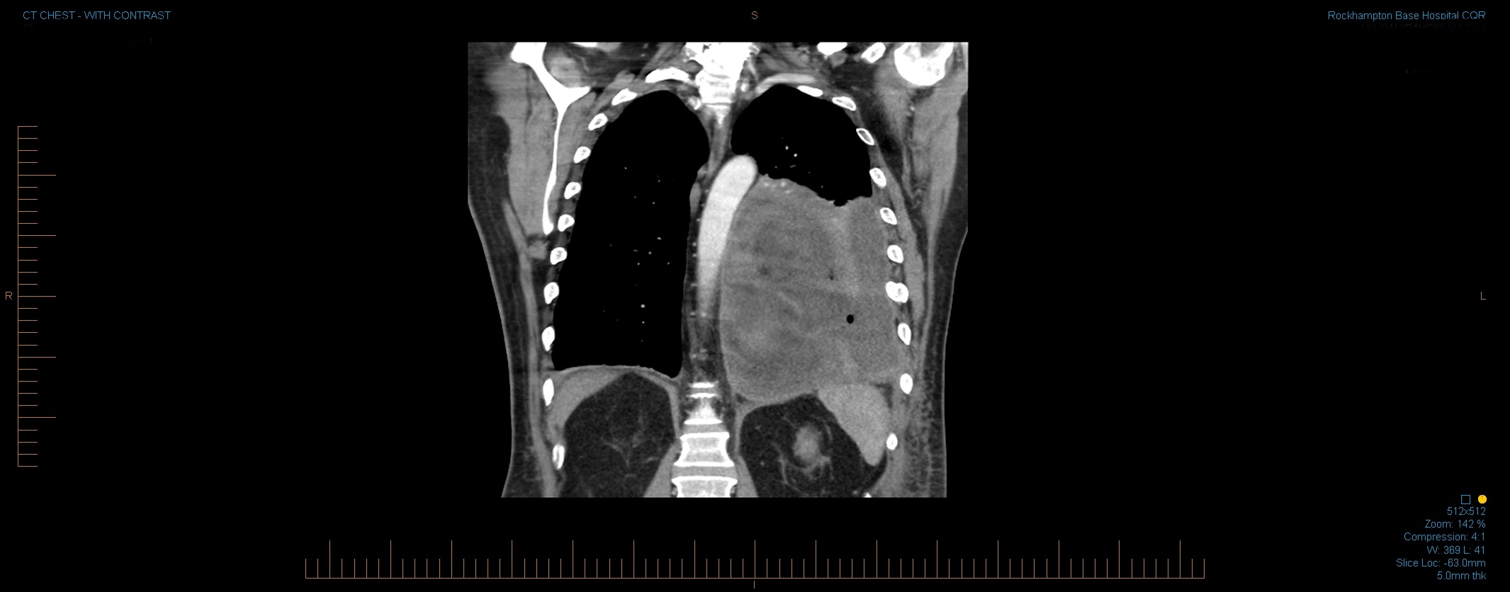
There is gas noted in the right side of the neck as well as the pneumomediastinum.
The lungs are clear. No effusion shown. Cardiac and mediastinal contours
are satisfactory. CT evaluation of the chest and neck could be considered if clinically indicated.
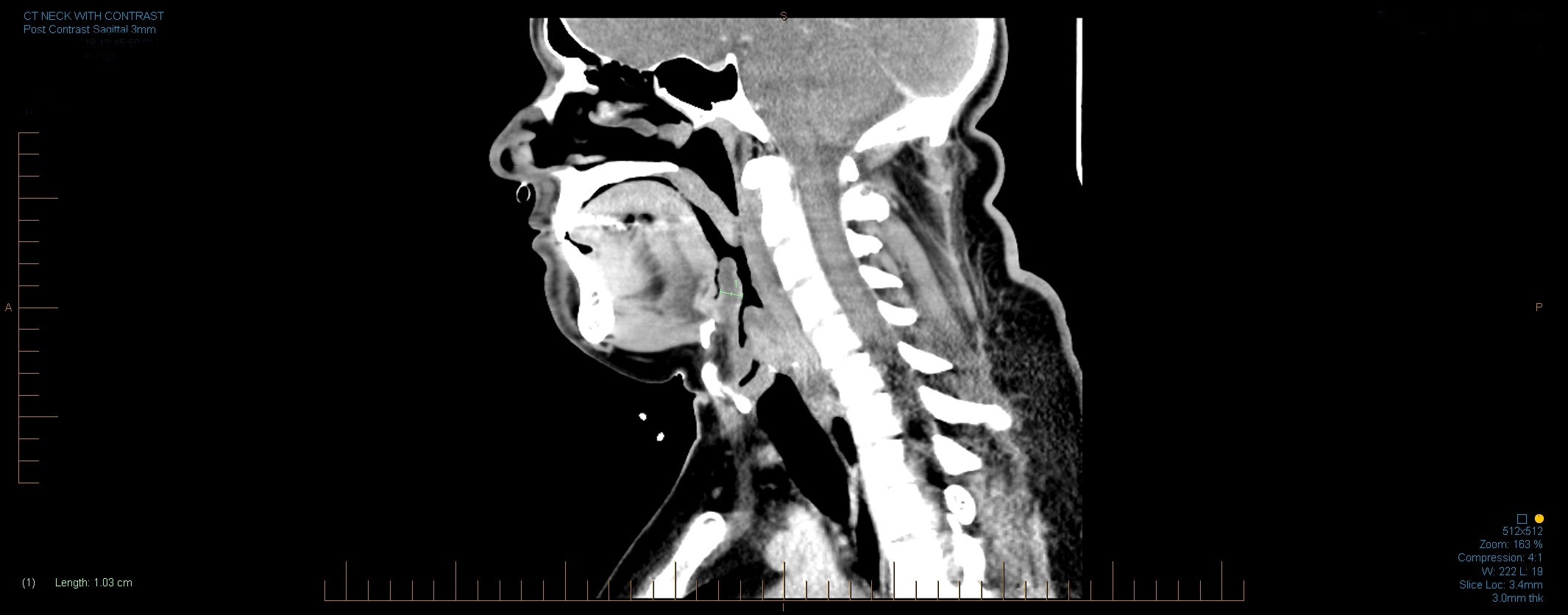
Inflammation of the epiglottis is known as epiglottitis. Epiglottitis is mainly caused by Haemophilus influenzae. A person with epiglottitis may have a fever, sore throat, difficulty swallowing, and difficulty breathing. For this reason, acute epiglottitis is considered a medical emergency, because of the risk of obstruction of the pharynx. Epiglottitis is often
Lung abscess is a type of liquefactive necrosis of the lung tissue and formation of cavities (more than 2 cm) containing necrotic debris or fluid caused by microbial infection.This pus-filled cavity is often caused by aspiration, which may occur during anesthesia, sedation, or unconsciousness from injury. Alcoholism is the most common condition predisposing to l
| « | previous | 1 | next | » |